A-PRF™ fibrin membrans for stimulation of the inflammatory cascade and faster healing

A-PRF™ | Advanced Platelet Rich Fibrin
Chairside access to the patients healing potential from venous blood
The concept of PRF (Platelet Rich Fibrin) is based on the centrifugation of whole blood without anticoagulants. (J. Choukroun et al. 2001). At the end of the spin, a fibrin clot containing the majority of the platelets and white blood cells is obtained.
This fibrin clot called Platelet Riche Fibrin or PRF will release gradually growth factors or cytokines in the site (VEGF, PDGF, TGF Beta, Thrombospondin)
The expected objective of these growth factors is to accelerate the soft tissue and bone healing.
A-PRF™ Clots in box
A-PRF™ | The process & application
A-PRF™ | Phlebotomy

Venous blood is drawn into A-PRF™ 10 ml tubes. This is done fast and each tube is placed into the centrifuge. Time is critical.
A-PRF™ | Spin

The A-PRF™ tubes are placed into the DUO centrifuge. It can hold 12 tubes. The protocol is pre-programmet and spin the tubes for 8 minutes. After the spin the lit opens and the next step can be performed.
A-PRF™ | Compression

The RBC are removed from the clot and the plasma clot is placed in the PRF box. A double lit is placed on top and in few minutes the exudate is removed from the clot and a each clot is turned into a fibrin membrane.
A-PRF | membrane

Each membrane is approximately 10 x 20 mm in size. If spinning 12 tubes an area of 14 – 20 cm2 may be covered with A-PRF™ membranes. The membrane size may differ from patient to patient.
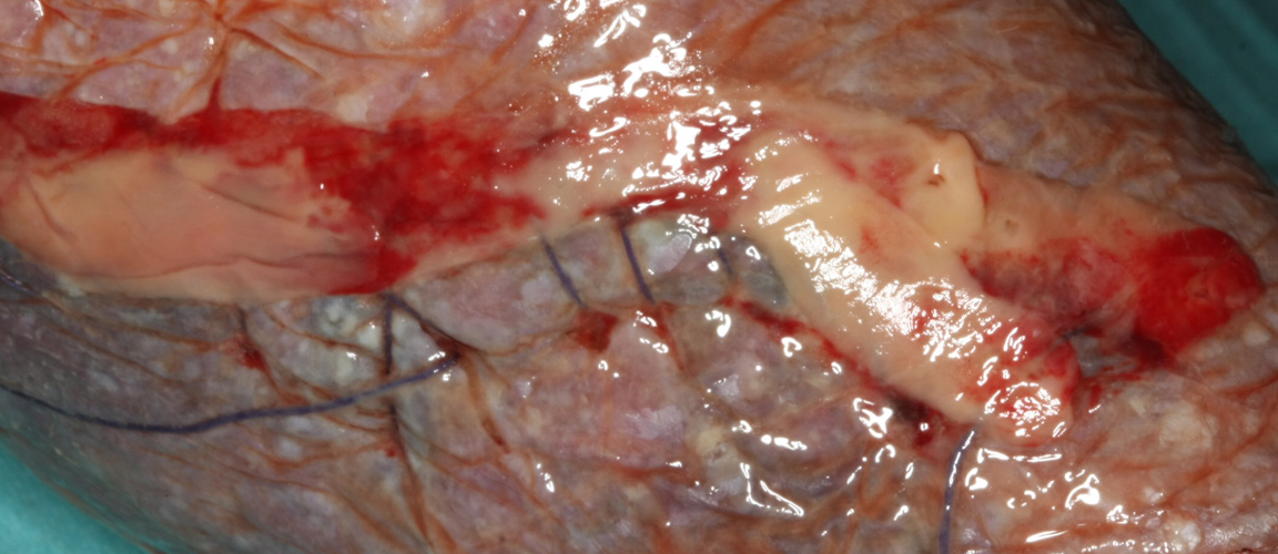
A-PRF | membrane – wound

Example of how A-PRF™ membranes cover a wound surface.
They stick to the surface easily. The picture to the right is 1 week later
A-PRF | membrane on wound after 1 week
The same A-PRF™ membranes as on the left picture – now one week later.
See more about wound healing here
A-PRF application | membranes cut into pieces

A-PRF™ membranes cut into small pieces ready to be mixed with biomaterials.
A-PRF application | membranes mixed with biomaterials

A-PRF™ membranes cut into small pieces and mixed with biomaterials and exudate enhance the bone formation process.
A-PRF application | membranes applied to bone graft

A-PRF™ membranes placed onto bone graft. Notice to thickness.
DUO Centrifuge solution
A-PRF™ | Videos
A-PRF | intro to PRF box
Short introduction creation of A-PRF™ membranes using the PRF box.
A-PRF | bone graft created from membranes and bonematerials
Bone particles mixed with A-PRF™ membrane cut into pieces formed into a graft. i-PRF™ is then injected onto the graft and it clots
A-PRF | sandwich graft
Combination of A-PRF™ and i-PRF™ in creating bone grafts from bone substituts.
A-PRF™ | Team and litteratur

Dr. Joseph Choukroun
Dr. Joseph Choukroun
received his diploma in 1979 in Montpellier, France. Specialist in anesthesiology in 1981, University of Montpellier. Fellowship of Pain Clinic, University of Strasbourg. Chief of staff of the Private Pain Clinic, Nice. President and creator of the SYFAC, international symposium on growth factors, Nice.
Inventor of the PRF technique. Author on several scientific and clinical papers for scientific journals. International Speaker.
International team
Dr. Joseph Choukroun is pushing the research on PRF together with scientists from the Laboratory of Clarion Research Group, Pennsylvania University (USA) and Repair-Lab, Institute of Pathology, Johannes Gutenberg University, Mainz (Germany) and Physicians around the world.
A-PRF™ box
with membranes ready to use – notice the instruments made for handling the membranes
PRFs can be regarded as dense fibrin biomaterial with bio-mechanical properties. A high density fibrin clot can serve as a biological healing matrix by supporting cell migration and cytokine release, expanding the range of its potential applications greatly.
Source:Classification of platelet concentrates: from pure platelet-rich plasma (P-PRP) to leucocyte- and platelet-rich fibrin (L-PRF), David M. Dohan Ehrenfest, Lars Rasmusson and Tomas Albrektsson, Department of Biomaterials, Institute of Clinical Sciences, The Sahlgrenska Academy at University of Gothenburg, Sweden
A-PRF™ | Advantages of PRF over PRP
- No biochemical handling of blood.
- Simplified and cost-effective process.
- Use of bovine thrombin and anticoagulants not required.
- Favorable healing due to slow polymerization.
- More efficient cell migration and proliferation.
- PRF has supportive effect on immune system.
- PRF helps in hemostasis.